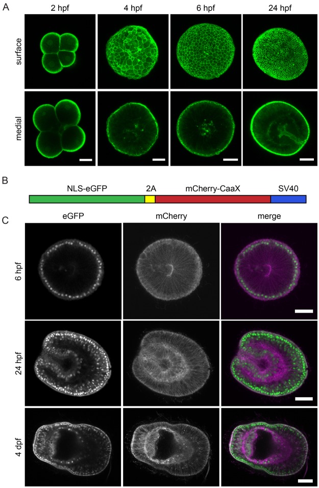
Figure 2.
Microinjection of recombinant Lifeact-eGFP protein and mRNA to fluorescently label nuclei and cell outlines. (A) Zygotes injected with ~3.4 mg/ml Lifeact-eGFP protein were fixed 2, 4, 6 and 24 hpf. Maximum projections of 5 z-planes near the surface or centre of the embryo are shown. Scale = 25 μm. (B) Schematic representation of bicistronic in vitro transcribed NLS-eGFP-V2A-mCherry-CaaX mRNA: eGFP with a nuclear localization signal (NLS) is coupled to mCherry with a C-terminal CaaX box for farnesylation and insertion into the membrane. The fluorophores are separated by the self-cleaving V2A peptide. (C) mRNA-injected embryos were fixed 6 or 24 hpf or 4 days post-fertilization (dpf). Left panels show eGFP-labeled nuclei, middle panels show mCherry-labeled membranes, and the right panel shows the merged images (eGFP green; mCherry magenta). Scale = 25 μm.

