
Figure 2.
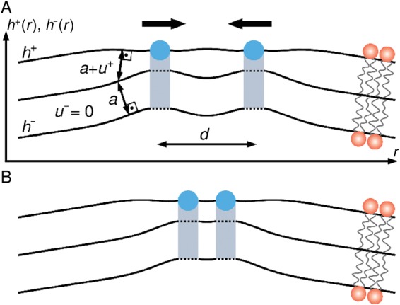
Schematic of bilayer-mediated interactions between protein wedges. (A) Amphipathic helices (viewed parallel to the helix axes and indicated in blue) deform the shape h+ and hydrophobic thickness u+ +a of the upper lipid bilayer leaflet, and may indirectly perturb h− and u− in the lower leaflet via the coupling between upper and lower leaflets. We set here u− = 0 (see main text). For simplicity, we assume in this schematic that the H0 helices are in the face-on orientation, and that the H0-induced membrane deformations are only a function of the spatial coordinate r measured perpendicular to the helix axes. Equation (3) predicts that, for large enough helix immersion depths, two protein wedges can be attracted to each other by bilayer-mediated wedge interactions (black arrows). (B) Our elastic model of bilayer-mediated wedge interactions predicts that, for large enough helix immersion depths, the distance d separating the axes of two neighboring amphipathic helices can take an optimal value set by the key lipid and protein properties captured by equation (3).