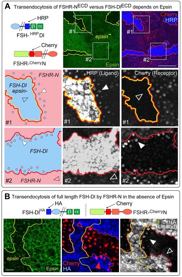
Figure 5. FSHR-N versus FSH-Dl transendocytosis depends on Epsin.
A, top) Transendocytosis of the extracellular FSH-Dl/FSHR-N bridge was assayed using HRP and Cherry extracellular tags, as in Figure 4, except that YFP-Rab5CA is expressed in both ligand and receptor expressing cells. Two independent types of clones were induced within the same disc. First, epsin¯ clones, outlined in yellow and marked “black” by the absence of Epsin (green, middle panel). Second, UAS>FSH-Dl clones (HRP, blue) generated by MAPS in a background of UAS>FSHR-N cells (Cherry, red), shown outlined in red (right panel). Some UAS>FSH-Dl clones are null for epsin (box #1); others are wildtype for epsin (box #2).
Box #1). FSH-Dl clone that is epsin¯ (blue, in the cartoon) in a background of FSHR-N cells (pink). Grey scale images of HRP and Cherry are shown in the middle and right panels. The FSH-Dl ectodomain accumulates in puncta in the abutting FSHR-N expressing cells (e.g., white arrowhead), whereas no accumulation of the FSHR-N ectodomain is detected in abutting FSH-Dl expressing cells (empty arrowhead).
Box #2). FSH-Dl expressing clone that is wildtype for epsin depicted and imaged as in the middle row. The results are reciprocal: the FSHR-N ectodomain accumulates in puncta in the neighboring FSH-Dl cells (e.g., white arrowhead right panel). In contrast, little or no accumulation of the FSH-Dl ectodomain is detected in puncta in the abutting FSHR-N cells (empty arrowhead, middle panel). Scale bar: 50μm.
B) FSH-Dl carrying an intracellular HA tag (FSH-DlHA) was used to monitor the fate of the Dl ICD (blue) following transendocytosis of the ligand from epsin¯ cells into FSHR-N receiving cells. As in A, two independent types of clones were induced within the same disc, namely, (i) epsin¯ clones (labelled as in A), and (ii) UAS>FSH-DlHA clones (blue) generated by MAPS in a background of UAS>FSHR-N cells (red). HA accumulation is apparent in puncta of FSHR-N cells that abut FSH-Dl epsin¯ cells (white arrowhead; grey scale image), but not in FSHR-N cells that abut wildtype FSH-Dl cells (empty arrow head). Taken together with the evidence of ligand transendocytosis in box #1 in (A), this indicates that the entire ligand has been internalized by the receiving cell. Concordant with the results shown in (A), transendocytosis of the receptor ectodomain in the opposite direction depends on whether the signal-sending cell is wildtype or mutant for epsin (middle panel): Cherry labeled puncta are evident in abutting FSH-Dl cells that retain epsin activity (red arrowhead), but are absent from FSH-Dl epsin¯ cells (empty red arrowhead). Scale bar: 10μm.

