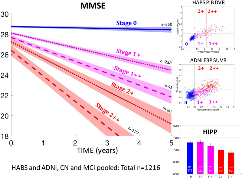
Fig. 3.
MMSE decline and hippocampal atrophy are more severe in individuals with high striatal Aβ than in individuals with very high cortical Aβ. Top and middle rows on the right: In both HABS and ADNI, individuals with high cortical Aβ are subdivided into four PET-Aβ substages, using both a striatal and a very high cortical Aβ threshold—1+: moderately high cortex, low striatum; 1++: very high cortex, low striatum; 21: moderately high cortex, high striatum; 2++: very high cortex, high striatum. Bottom right: hippocampal volumes by PET-Aβ substages in nondemented older adults. Striatal PET-Aβ, but not very high cortical PET-Aβ, is associated with lower hippocampal volumes. Left: MMSE decline by PET-Aβ substages in nondemented older adults. Groups with high striatal Aβ (2+ and 2++) demonstrated the fastest decline. Error bars are 95% confidence intervals. See the last row of Table 3 for statistics. Abbreviations: Aβ, amyloid β; ADNI, Alzheimer’s Disease Neuroimaging Initiative; HABS, Harvard Aging Brain study.

