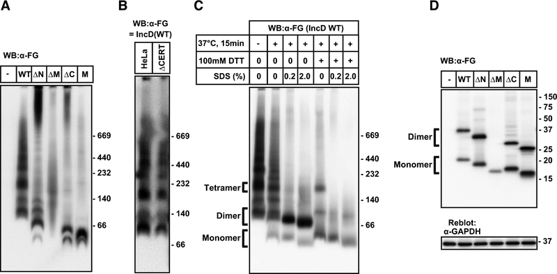
Figure 4. IncD forms higher order oligomers by dimers.
(A) HeLa cells were transfected with pcDNA4/TO encoding FG-IncD WT, FG-IncD ΔN, FG-IncD ΔM, FG-IncD ΔC, FG-IncD M, or an empty vector. Cell extracts were prepared from the cells with 1% dodecylmaltoside-containing lysis buffer. The cell extracts were separated by Blue-Native-PAGE as manufacturer’s instruction and analysed by Western blotting with anti-FLAG antibody. (B) HeLa cells or CERT-deficient HeLa cells were transfected with plasmid encoding FG-IncD WT and cell extracts were prepared as described above. The cell extracts were separated by Blue-Native-PAGE and analysed by Western blotting with anti-FLAG antibody. (C) HeLa cells were transfected with pcDNA4/TO encoding FG-IncD WT and cell extracts were prepared as described. For samples of Blue-Native-PAGE, cell extracts were incubated at 37 °C for 15 min in the presence or absence of the indicated reagents. (D) The cells described in Fig. 4A were lysed by LDS-containing nonreducing loading buffer. The lysate was subjected to SDS-PAGE under non-reducing conditions and, after electrophoresis, IncD constructs were detected by Western blotting. The membranes were reblotted with an anti-GAPDH antibody.

