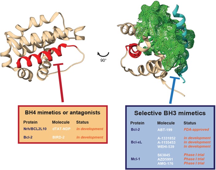
Figure 1. Differential targeting of distinct pro-survival Bcl-2 protein domains.
Representation of a typical Bcl-2 protein structure (here Nrh/Bcl2L10/Bcl-B, PDB: 4B4S). In red, the N-terminal BH4 helix. In blue, the BH3 alpha helix of Bim bound to Nrh. In green (mesh), the surface of the hydrophobic BH3 binding pocket of Nrh. The different selective molecules for the separate targeting of pro-survival Bcl-2 protein BH4 domains (BH4 mimetics or antagonists) or hydrophobic BH3 binding pocket (BH3 mimetics) are listed in the tables.

