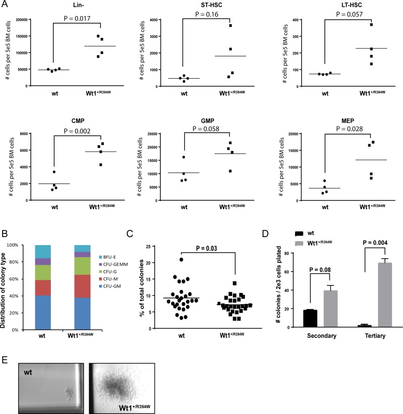
Figure 2. In vitro progenitor cell analysis of 2-month old wild type (wt) and Wt1+/R394W bone marrow.
(A) Absolute number of cells in each progenitor cell compartment per 5 × 10⁵ live bone marrow cells of wt or Wt1+/R394W mice (n = 4 each), as analyzed by flow cytometry. Short-term hematopoietic stem cells (ST-HSCs) were defined as lineage (Lin)-sca1+ckit+ (LSK) and CD34+135–; long-term (LT)-HSCs as LSK and CD34–135–; common myeloid progenitors (CMP) as Lin-sca-ckit+CD34+FcγR-; granulocyte-monocyte progenitors (GMP) as Lin-sca-ckit+CD34+FcγR+; and megakaryocyte-erythroid progenitors (MEP) as Lin-sca-ckit+CD34-FcγR–. (B) Distribution of colony type formation in methylcellulose culture at Day 7 after initial plating. Lineage-depleted bone marrow cells from 2-month old mice were originally plated at 2 × 10³ cells per mL of methylcellulose, in triplicate. Results are representative of nine separate experiments. BFU-E = burst-forming unit-erythroid; CFU-GEMM = colony-forming unit-granulocyte, erythrocyte, monocyte/macrophage, megakaryocyte; CFU-G = colony-forming unit-granulocyte; CFU-M = colony-forming unit-macrophage; CFU-GM = colony-forming unit-granulocyte/macrophage. (C) Number of BFU-E colonies generated from Wt1+/R394W lineage-depleted bone marrow compared to wt bone marrow at Day 7. (D) Colony counts after secondary and tertiary re-plating of Wt1+/R394W or wt bone marrow cells in methylcellulose culture, done in triplicate. Cells were harvested and re-plated every 10–12 days. (E) Representative colony size at tertiary re-plating (magnification 20×). Horizontal bars represent the mean values, error bars represent the SEM.

