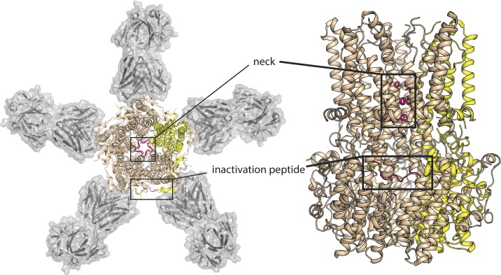
Figure 1.
Structure of chicken BEST1 with neck and inactivation peptide highlighted. Two views of chicken BEST1 (PDB ID 4RDQ): at left, perpendicular to the membrane, and at right, parallel to the membrane. In each cartoon, four BEST1 subunits are colored in wheat, with one colored yellow for emphasis. The sidechains that define the neck (I76, I80, I84) and the inactivation peptide (356RPSFLGS362) from the yellow subunit are highlighted in hot pink. In the top-down view, Fab fragments 10D10 are shown with gray surface rendering.

