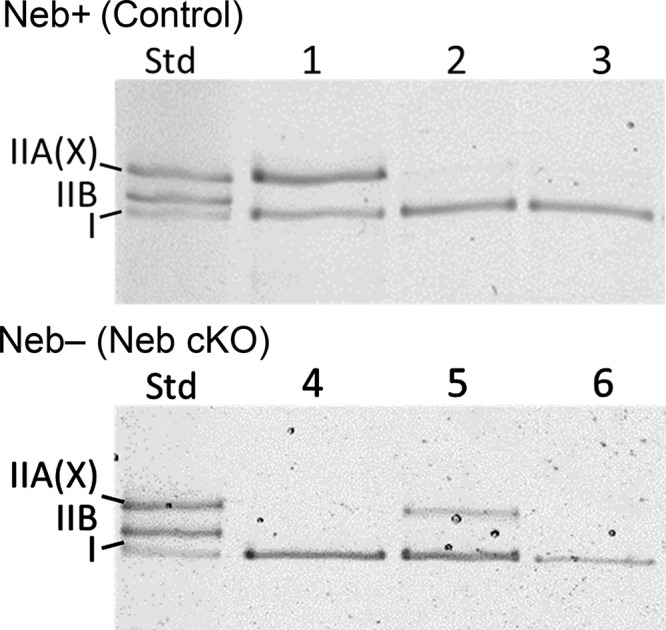
Figure 1.

MHC isoform expression analysis with SDS-PAGE. MHC isoform composition of fiber bundles dissected from skinned soleus muscle from Neb-expressing control muscle (top) and Neb-deficient muscle (bottom). The left lane is a standard (Std) that was made by mixing soleus and tibialis cranialis muscle lysate, revealing type I, IIB and IIA(X) MHC bands; note that this electrophoresis system is unable to separate IIA and IIX MHC isoforms. Lanes 1 and 5 are examples of bundles that contained both type I and IIA(X) MHC; these were not used in the present study. Lanes 2, 3, 4, and 6 contain solely type I MHC, and they are examples of fiber bundles that were entered in the present work.
