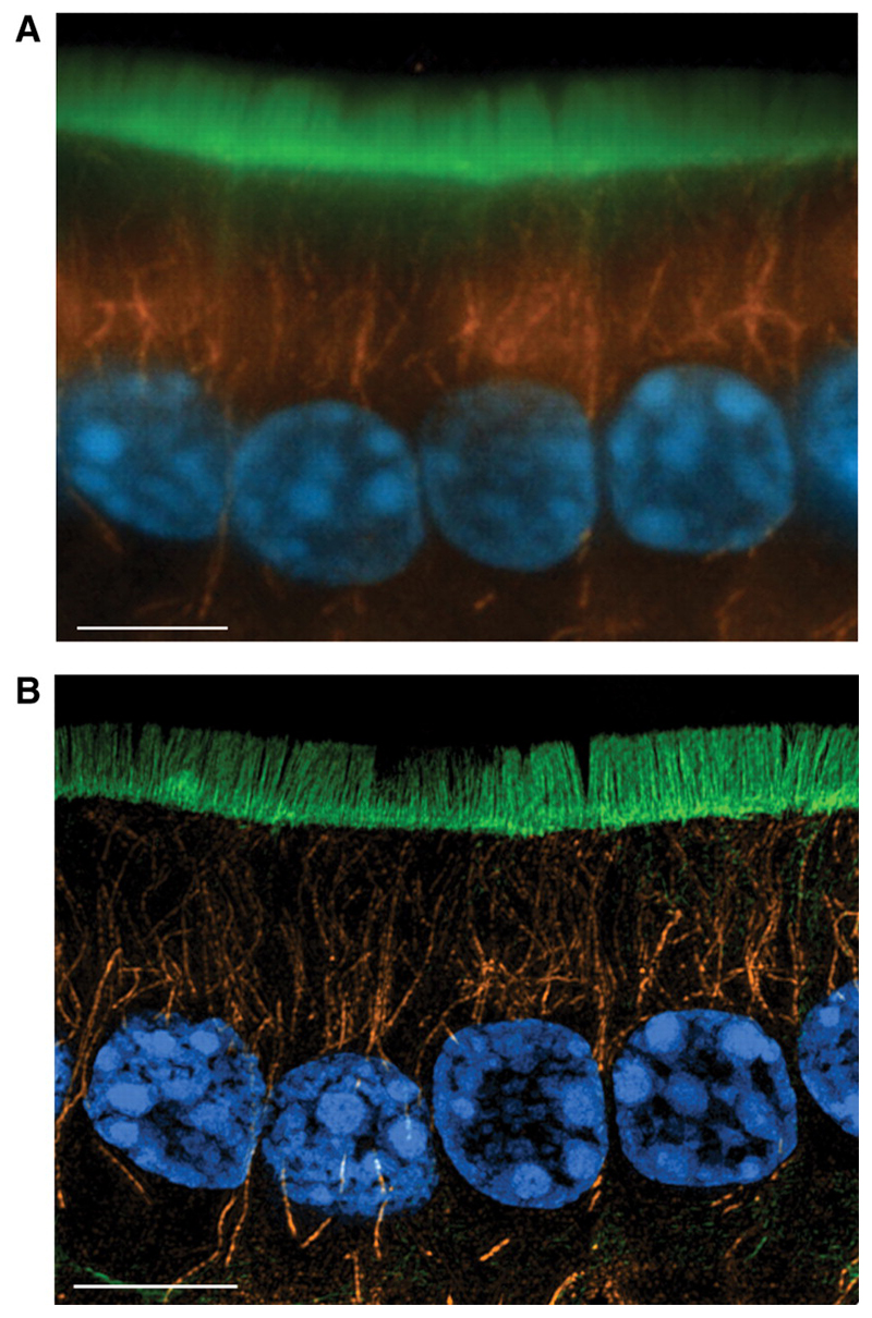
Figure 6. 3DSIM imaging on OMX—cryosections.
10μm cryosection of formaldehyde fixed mouse small intestine, stained with DAPI to show nuclei (blue), anti-tubulin/Alexa Fluor 568 to show microtubules (orange) and FITC-phalloidin to show F-actin (green), (a) deconvolved conventional widefield image, 5 z-section maximum intensity projection, (b) image cquired using the 3DSIM protocol on OMX v2. Wavelengths were acquired sequentially, reconstructed then aligned and fused. 5 z-section maximum intensity projection. Scale bar represents 5μm. Emma King and Paul Appleton (University of Dundee).

