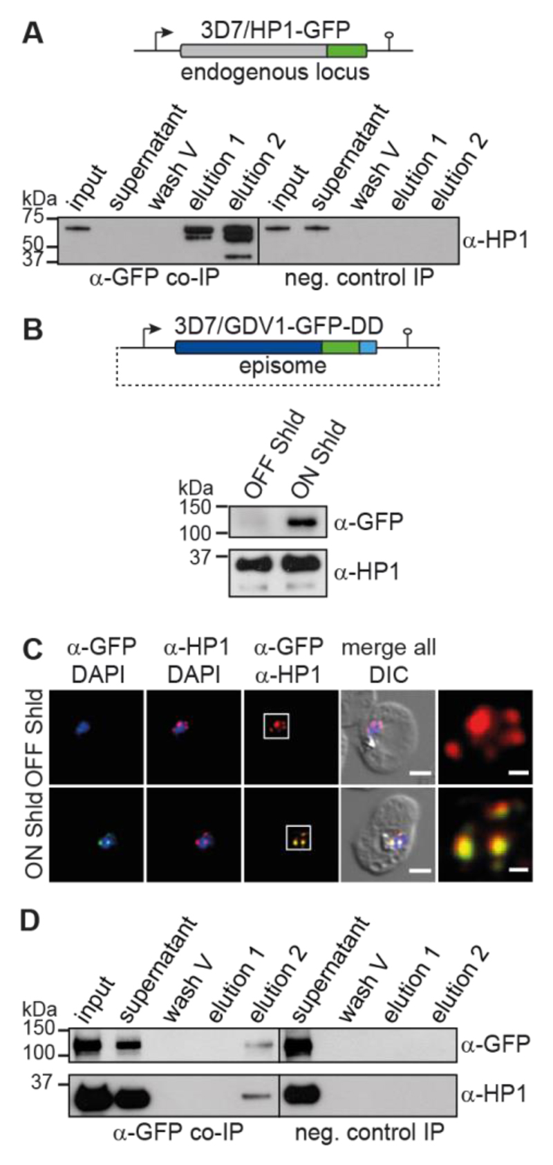
Fig. 1. GDV1 interacts with HP1.
(A) Endogenous hp1 locus in 3D7/HP1-GFP parasites and α-HP1 Western blots of the α-HP1-GFP co-IP and negative control samples. Results are representative of three biological replicates. (B) gdv1-gfp-dd expression plasmid and α-GFP Western blots of 3D7/GDV1-GFP-DDOFF and 3D7/GDV1-GFP-DDON parasites. α-HP1 antibodies served as loading control. (C) GDV1-GFP-DD/HP1 co-localisation IFAs in 3D7/GDV1-GFP-DDOFF and 3D7/GDV1-GFP-DDON trophozoites (24-32 hpi). DIC, differential interference contrast. Scale bar, 2.5 μm (0.5 μm for the magnified views in the rightmost images). Results are representative of three biological replicates. (D) α-GFP and α-HP1 Western blots of the α-GDV1-GFP-DD co-IP and negative control samples. Results are representative of three biological replicates.

