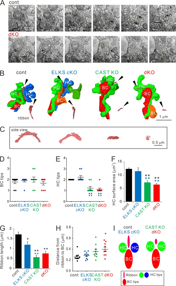
Figure 4.
3D reconstruction of rod terminals in the OPL shows morphological abnormalities in CAST KO and dKO retinae. (A) Representative FIB-SEM images from control and dKO (300-nm interval). The rod terminals indicated by yellow lines were 3D-reconstructed in A. Arrows indicate synaptic ribbons. (B and C) Representative images of 3D-reconstructed triads were acquired by FIB-SEM. Small truncated ribbons (pink bands indicated by black arrowheads) occur in a single HC tip in the CAST KO and dKO. HC (green, blue), BC (red, orange). Representative examples of ribbons are illustrated (as viewed from the bottom; black arrowhead); ribbon profiles are demonstrated in C. (D–F) Analysis of BC and HC tips inserted into rod terminals. Consistent with the reduced HC tip numbers (E) in CAST KO and dKO retinae, the respective accumulated HC surface areas (F) are also significantly decreased. Colored asterisks indicate significant differences compared with the respective colored genotypes, mean ± SEM; n = 9. **, P < 0.01 (one-way ANOVA, post hoc Tukey test). (G) The entire ribbon length in each terminal was significantly decreased in all three deletion mutants compared with control. (H) The shortest distance from the ribbon to the BC tip is significantly increased in the dKO, mean ± SEM; n = 9. *, P < 0.05; **, P < 0.01 (one-way ANOVA, post hoc Tukey test). (I) Simplified schematic models of triad complexes composed by a ribbon, two HCs, and one or two BCs. In CAST KO and dKO retinae, the two HC tips switch to a branched single tip with reduced ribbon length.

