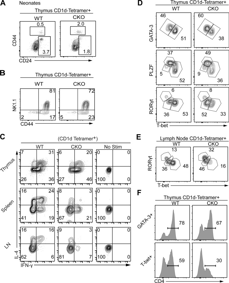
Figure 3.
Remnants of iNKT cells in Sox4 CKO mice are selectively depleted of NKT1 cells. (A) Representative flow cytometric analyses show a proportional enrichment for DP (CD8α+) CD1d-tetramer+ cells in neonatal CKO mice. Note that a general trend for decreased CD24 expression in Sox4 CKO DP thymocytes obscures the CD24+ NKT0 cells. (B) iNKT maturation marker CD44 and NK1.1 were analyzed in thymic iNKT cells of WT and CKO mice. Representative profiles from five independent experiments. (C) Programmed production of IL-4 and IFNγ from remnants of Sox4 CKO iNKT cells from indicated tissues was compared with normal iNKT cells (with no stimulation profiles). Representative profiles from four independent experiments. (D and E) iNKT effector subsets identified by the expression of indicated TFs in iNKT cells show decreased production of T-bet+ NKT1 cells in remnants of Sox4 CKO iNKT cells. Frequencies of each subset were assessed in the thymus (D) and peripheral LNs (E). Representative profiles from three independent experiments. (F) Proportions of CD4 iNKT cells within thymic GATA3+ or T-bet+ iNKT cells are shown. Representative plots from three independent experiments.

