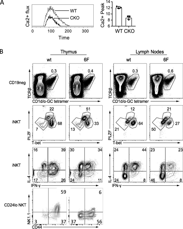
Figure 5.
Evidence for impaired TCR signaling in Sox4-deficient thymocytes. (A) Impaired Ca2+ signaling in thymocytes lacking SOX4. Left, kinetic analysis of Ca2+ flux (ratios of Fluo 3:Fura red) in DP thymocytes from WT and Sox4 CKO mice after stimulation with CD3ε cross-linking. Representative plots from four independent experiments. Right, averages of peak flux (n = 4/genotype). Error bars denote SD. (B) Thymocytes from Cd2476F/6F (6F) mice with compromised TCR-signaling capacity also show impaired NKT1 cell generation and function. Shown from top to bottom panels are iNKT cell frequencies, iNKT cell subset distributions, effector cytokine production, and iNKT cell maturation profiles in the thymus and LNs of WT and Cd2476F/6F mice. Representative plots from three independent experiments using mice of age ranging from 4 to 6 wk old (thymus) or 8 to 12 wk old (LN).

