Figure 1.
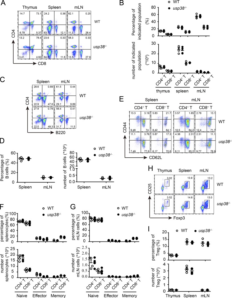
USP38 deficiency does not affect homeostatic immune cell development. All the experiments below were carried on 6–10-wk-old littermates. (A and B) Cytoflow analysis (A) and quantification (B) of CD4- or CD8-positive populations in the indicated tissues out of usp38−/− or WT mice. (C and D) Cytoflow analysis (C) and quantification (D) of B cells in the indicated tissues out of usp38−/− or WT mice. (E–G) Cytoflow analysis (E) and quantification (F and G) of naive (CD62L+CD44−), effector (CD62L−CD44+), and memory (CD62L+CD44+) CD4+ T cell populations, as well as CD8+ T cells, in the indicated tissues out of usp38−/− or WT mice. (H and I) Cytoflow analysis (H) and quantification (I) of T reg cells in the indicated tissues out of usp38−/− or WT mice. Data are representative of four (A–I) independent experiments. Statistical significance was determined by Student’s t test. Error bars indicate the mean ± SEM.

