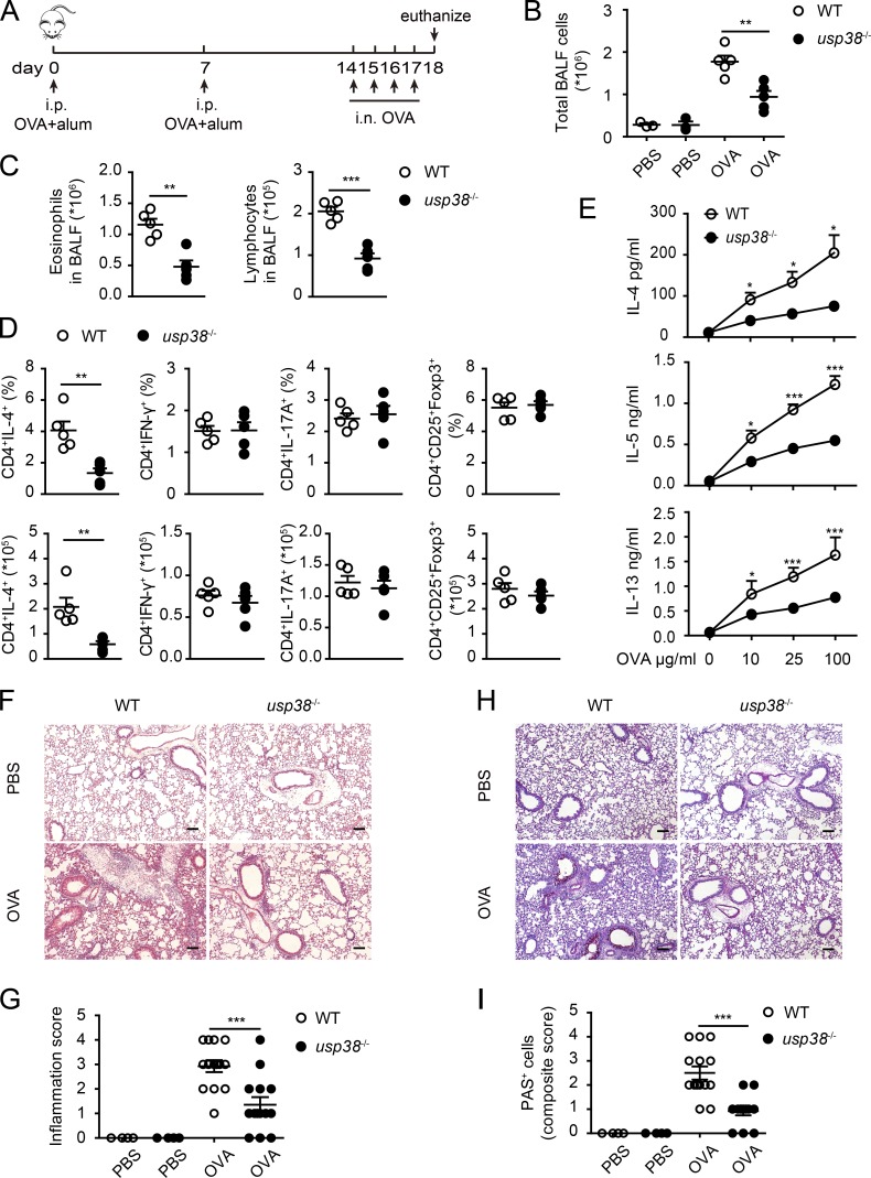
Figure 2.
USP38 critically promotes OVA-induced asthma. (A–I) 6–8-wk-old female usp38−/− mice and their littermate controls (WT) were analyzed individually. PBS group (n = 3); OVA group (n = 5). (A) A schematic diagram of OVA-induced allergic asthma. (B) Total number of BALF cells. (C) Numbers of eosinophils or lymphocytes in BALF by flow analysis. (D) Percentages and absolute numbers of Th1 (CD4+IFN-γ+), Th2 (CD4+IL-4+), Th17 (CD4+IL-17A+), and T reg (CD4+CD25+Foxp3+) cells in mediastinal lymph nodes (medLNs) from OVA-treated mice. (E) Levels of Th2 cytokines determined by ELISA from culture supernatants of medLNs cells treated with the indicated concentrations of OVA protein for 72 h. (F) H&E staining of lung sections from PBS- or OVA-treated mice. Original magnification is 10×. Bars, 100 µm. (G) Pathological score of perivascular and peribronchiolar inflammation as shown in F. (H) PAS staining of lung sections from PBS- or OVA-treated mice. Original magnification is 10×. Bars, 100 µm. (I) Quantification of goblet cell hyperplasia as shown in H. Data are representative of four (A–C) or two (D–I) independent experiments. Statistical significance was determined by Student’s t test; *, P < 0.05; **, P < 0.01; ***, P < 0.001. Error bars indicate the mean ± SEM.

