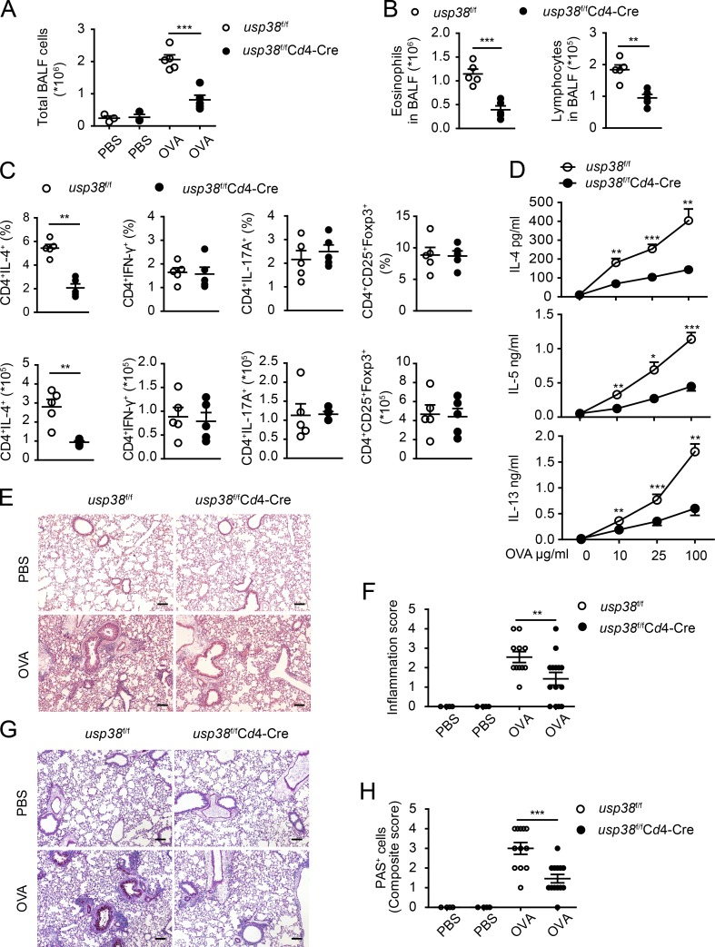
Figure 4.
T cell–derived USP38 mediates the pathogenesis of OVA-induced asthma. (A–H) Mice with T cell–specific deletion of usp38 (usp38f/fCd4-Cre) and their littermate controls (usp38f/f) were used for OVA-induced asthma model. PBS group (n = 3); OVA group (n = 5). (A) Total number of BALF cells. (B) Quantification of eosinophils or lymphocytes in BALF determined by flow analysis. (C) Percentages and absolute numbers of the indicated T cell subpopulations in medLNs. (D) Levels of Th2 cytokines determined by ELISA from Th2 ex vivo recall response. (E) H&E staining of lung sections from PBS- or OVA-treated mice. Original magnification is 10×. Bar, 100 µm. (F) Pathological score of perivascular and peribronchiolar inflammation as shown in E. (G) PAS staining of lung sections from PBS- or OVA-treated mice. Original magnification is 10×. Bars, 100 µm. (H) Quantification of goblet cell hyperplasia as shown in H. Data are representative of four (A and B) or two (C–H) independent experiments. Statistical significance was determined by Student’s t test; *, P < 0.05; **, P < 0.01; ***, P < 0.001. Error bars indicate the mean ± SEM.

