Figure 3.
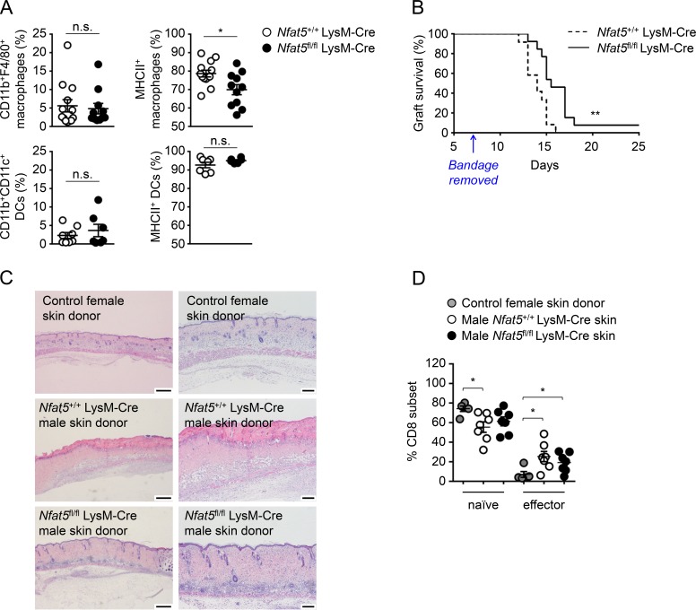
Rejection of myeloid-specific NFAT5-deficient skin transplants. (A) Percentage of macrophages (CD11b+ F4/80+) and DCs (CD11b+ CD11c+) and their MHCII expression in skin biopsies of Nfat5+/+ LysM-Cre (wild-type) and Nfat5fl/fl LysM-Cre mice. Statistical significance was determined by an unpaired t test. *, P < 0.05. n.s., not significant. (B) Survival of skin grafts from Nfat5+/+ LysM-Cre and Nfat5fl/fl LysM-Cre male donor mice transplanted in wild-type female recipients. Results correspond to 10 independent transplant experiments, in each of which separate recipient female mice were respectively transplanted with skin from Nfat5+/+ LysM-Cre (wild-type) and Nfat5fl/fl LysM-Cre male mice. Transplant rejection was monitored after day 7, when the protective postsurgery bandage was removed (indicated by the arrow). Median survival for skin grafts of Nfat5+/+ LysM-Cre male mice (n = 12) was 14 d, and median survival for skin grafts of Nfat5fl/fl LysM-Cre male mice (n = 13) was 15.5 d (see Fig. S2 for representative pictures illustrating the time course of graft rejection). P = 0.0023, calculated with a Mantel–Cox log-rank test. (C) Histological analysis (hematoxylin and eosin staining) of skin grafts from a female wild-type donor (as quality control for the surgical procedure) and male Nfat5+/+ LysM-Cre and Nfat5fl/fl LysM-Cre donors 10 d after transplant in female recipients. Photographs are representative of histopathology analyses done in four control female skin transplants, six Nfat5+/+ LysM-Cre male skin transplants, and five Nfat5fl/fl LysM-Cre male skin transplants. Scale bar is 500 µm for the photographs in the left column, and 200 µm for the enlarged images in the right column. (D) Proportion of naive and effector CD8+ T cells in the spleens of transplanted mice. CD8+ T cells were analyzed in five independent transplant experiments, four of which included parallel controls with female mice transplanted with skin of a wild-type female donor (as shown in C). Recipient mice were sacrificed on the day when clear rejection was observed for wild-type Nfat5+/+ LysM-Cre male skin grafts (between days 12 and 16 after transplant). Results in the graphics are the mean ± SEM. *, P < 0.05. Significance was determined by a Mann–Whitney test.

