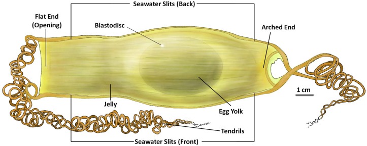
Fig 1. External features of the S. stellaris egg case at stage 1.
The internal ellipsoid egg yolk and blastodisc visible at stage 1 of development. The egg case membrane was filled with jelly surrounding the yolk. There were four seawater slits located at each corner of the egg case, two can be seen from the front (as shown in Fig 1), and another two at the back of the egg case (not shown). The flat end of the egg case will be the site of opening during hatching, whereas the arched end remains firm and closed during hatching. There are four tendrils attached at each corner of the egg case. The key feature for stage 1 was no visible embryo on top of the egg yolk. See S1 File for original photographs of Fig 1 illustration.

