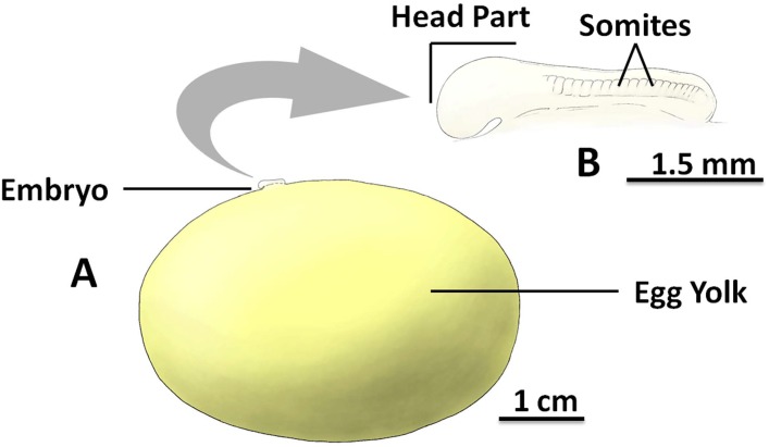
Fig 2. The inside of the S. stellaris egg case at stage 2.
A: The egg yolk mass and the associated embryo to scale. B: The magnified embryo with indication of key morphological features (lateral view). The embryo consists an anterior (head and trunk primordia) and posterior (tail bud), where the somites (segmented mesoderm) can be found. The key feature for stage 2 was the visible embryo developed on top of the egg yolk membrane without a long tail. See S2 File for original photographs of Fig 2 illustrations.

