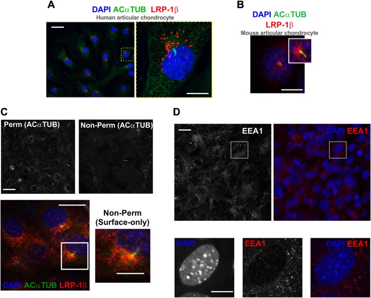
Figure 4.
Membranous hot spot of LRP-1β associated with primary cilium. A) Immunofluorescent staining of primary cilia (green) and LRP-1β (red) in human articular chondrocytes. B) Immunofluorescent staining of LRP-1β (red) and primary cilia (green) in wild-type (WT) mouse chondrocytes. C) Immunofluorescence images shows surface-only staining of LRP-1β (red) in WT cells that concentrated at the base of the primary cilium (green) in cells without permeabilization as validated by the absence of a majority of acetylated α-tubulin signal (black and white images above). D) Immunofluorescence of EEA-1 (red) in mouse WT chondrocytes. Nuclei were labeled with DAPI. Scale bars, 10 µm.

