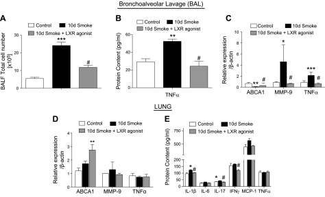
Figure 5.
Effect of LXR activation on cigarette smoke–induced acute pulmonary inflammation and MMP activation. Total inflammatory cell counts in BAL fluid (BALF) of mice exposed to cigarette smoke for 10 d with or without LXR agonist treatment vs. room air–exposed control mice (n = 4). A) Protein concentration of TNF-α. B) mRNA analysis of ABCA1, MMP-9, and TNF-α in BAL cells of mice exposed to cigarette smoke for 10 d with or without LXR agonist treatment vs. room air–exposed control mice. C) mRNA analysis of ABCA1, MMP-9, and TNF-α in the lungs of mice exposed to cigarette smoke for 10 d with or without LXR agonist treatment vs. room air–exposed control mice. D) Protein concentrations of IL-1β, IL-6, IL-17, IFN-γ, monocyte chemoattractant protein 1 MCP-1, and TNF-α measured by Luminex cytokines array system in the lungs of mice exposed to cigarette smoke for 10 d with or without LXR agonist treatment vs. room air–exposed control mice. E) β-Actin was used as a housekeeping gene control for real-time PCR. *P < 0.05, **P < 0.01, ***P < 0.001 when compared with controls, #P < 0.05 compared with 10 d of cigarette smoke exposure (significantly significant).

