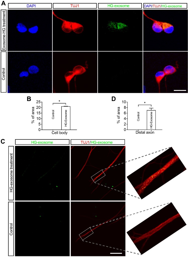
Figure 2.
Cell bodies and distal axons of DRG neurons internalize HG exosomes. A, C) Representative immunofluorescent images acquired from the cell body compartment show the presence of HG exosomes (green) within the cytoplasm of TUJ1+ DRG neurons (A, red) or TUJ1+ distal axon (C, green), when Exo-fect labeled HG exosomes were applied to the cell body compartment or the axonal compartment only, respectively. The nuclei were labeled with DAPI (blue). B, D) Quantitative data of green fluorescent signals of labeled HG exosomes within cell bodies and distal axons (n = 3 chambers/group). Scale bars: 10 μm (A), 5 μm (C). *P < 0.05.

