Figure 1.
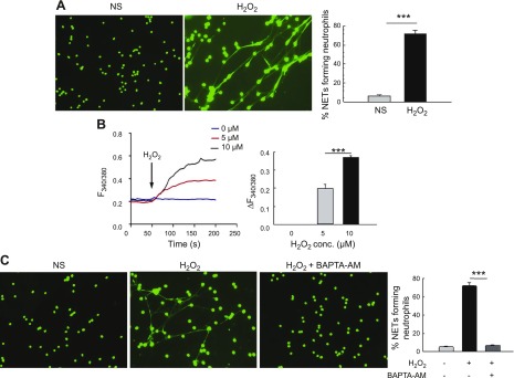
A) Exogenous H2O2 induces robust NET formation and calcium influx in vitro in neutrophils. Representative fluorescence images of WT neutrophils unstimulated (NS) or stimulated with H2O2 for 4 h. Cells were fixed and NETs were stained with DNA dye Sytox Green. Original magnification, ×200. Percentage of NET-forming neutrophils (mean ± se) from 4 independent experiments is shown in the bar graph. B) Calcium imaging by Fura-2 acetoxymethyl ester fluorescence measurement was performed on primary neutrophils in the absence (NS) or presence of 0–10 µM H2O2. Average analog plots of the fluorescence ratio (340/380 nm) from an average of 40–50 cells are shown. The bar graph shows the average data from 3 independent experiments for changes in cytosolic calcium under these conditions in unstimulated and H2O2-stimulated primary neutrophils. C) Neutrophils were pretreated for 30 min with 5 µM BAPTA-AM before stimulation with 10 mM H2O2 for 4 h. Representative fluorescent images of neutrophils are shown (original magnification, ×200). Bar graph shows quantification (average ± se) of NET-forming neutrophils from 3 independent experiments. ***P < 0.001.

