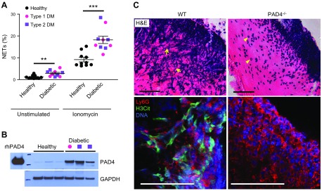
Figure 4.
Enhanced NETosis in neutrophils from patients with diabetes and deposition of NETs in mouse skin wounds. A) Both T1DM and T2DM prime human neutrophils to produce NETs. B) PAD4 protein expression is strikingly up-regulated in the neutrophils of patients with diabetes. T1DM, pink circle; T2DM, purple square. C) Hematoxylin and eosin staining revealed prominent extracellular DNA structures, appearing as blue streaks (arrows), in the scab of WT mice 3 d postinjury (top). Neutrophils remain intact (arrowheads) and NET structures are absent in the scab of PAD4−/− mice. Confocal immunofluorescence microscopy showed that H3Cit (green) colocalizes with externalized DNA (blue) in neutrophil (Ly6G) -rich area (red) in WT, but not PAD4−/− scab, which indicates the crucial role of PAD4 in NETosis in the mouse skin wounds (bottom). Scale bars, 50 μm. Adapted from Wong et al. (95).

