Figure 1.
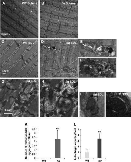
Lipin-1 deficiency results in mitochondrial aggregation and an increase in autophagic vacuoles. Representative electron micrographs of soleus of WT (A) and fld (B), and EDL of WT (C) and fld (D–J) mice. The morphology of some of the vacuoles resembles autophagosomes (E, F, yellow arrows) and autolysosomes (E, blue arrows). G–J) Representative micrographs showing mitochondrion-containing autophagic vacuoles. K) Quantification of the number of mitochondrial aggregates per 100 μm2. L) Quantification of the number of autophagic vacuoles per view field. **P < 0.01.

