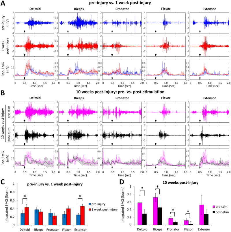
Figure 3: Effects of cervical electrical stimulation on forelimb muscle synergies post-injury.
(A) Raw EMG signals from the forelimb muscles during reaching and grasping pre-injury (blue trace) and 1 week post-injury (red trace), and the mean (±SD, color shading) rectified (Rec.) EMG signals (n = 20 trials each) from the same rat. Black arrow indicates the initiation of lifting the forelimb paw. (B) Raw EMG signals from the forelimb muscles during reaching and grasping at 10 weeks post-injury pre-stimulation (pink traces) and post-stimulation (black traces), and the mean (±SD, color shading) rectified EMG signals (n = 20 trials each) from the same rat. Black arrow indicates the initiation of lifting the forelimb paw. (C) Comparison of the mean (±SEM) integrated EMG values (n = 40 trials, 5 rats) obtained pre-injury and 1 week post-injury during reaching and grasping without stimulation. Two muscles, i.e., the deltoid and extensor, had significantly higher EMG activity levels 1-week post-injury compared to pre-injury when performing the task. (D) Comparison of the mean (±SEM) integrated EMG values (n = 40 trials, 5 rats) obtained prior (pre-stim) and immediate after (post-stim) receiving epidural stimulation at 10 weeks post-injury. *: significantly different at p < 0.05

