
Figure 10. Helminthic induction and maintenance of TGFβ production requires IL4 production by Foxp3− CD4 T cells.
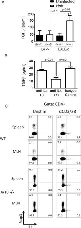
(A) TGFβ concentration in supernatants of anti-CD3/28-stimulated MLN cultures from Hpb-infected and uninfected 8–9 week old male IL4−/− or WT BALB/c mice, as measured by ELISA. Data show mean±SD from ≥3 independent experiments, with each experiment containing multiple determinations (N indicates the number of independent determinations). p value as indicated on the figure between Hpb-infected vs. uninfected groups; differences between groups determined by unpaired Welch’s t-test. (B) Anti-CD3/28 stimulated MLN T cells from Hpb-infected WT BALB/c mice, as described in Methods were cultured with anti-IL4 blocking (anti-IL4 (+)) isotype control antibodies (Isotype Control), or no antibody added (anti-IL4(–)), as indicated. Supernatants were analyzed for TGFβ content by ELISA. Data show mean±SD from a representative experiment of 5 independent experiments, with each experiment containing multiple determinations; p values as shown between groups; differences between groups determined by unpaired Welch’s t-test. (C) Representative dot plots of anti-CD3/28-stimulated splenocyte and MLN cultures from Hpb-infected 8–9 week old male WT BALB/c or Jα18−/− mice, with Brefeldin A added to cultures for the last 12 hours. Cells were stained for CD4, Foxp3 and IL4 using Foxp3 staining protocol. Data is representative example of 3 independent experiments for each group.