
Figure 11. GATA3 is expressed on Foxp3− CD4 T cells and Foxp3+ CD4 Tregs during GVHD and is critical to IL4 and TGFβ production by T cells.
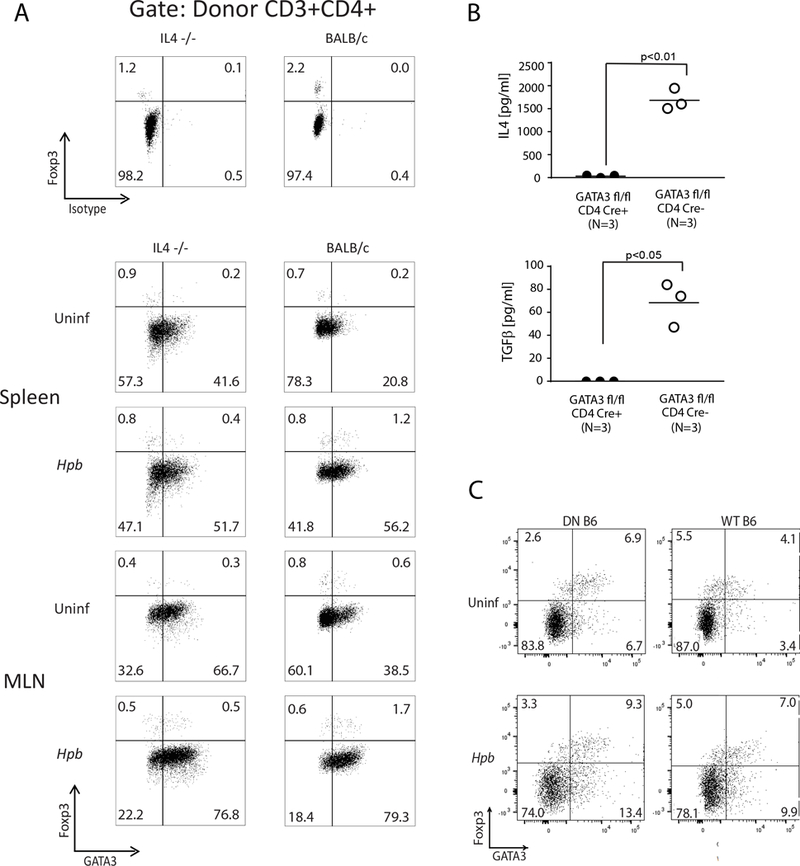
(A) Representative dot plots from spleen and MLN cells isolated from uninfected and Hpb-infected IL4−/− (left) or WT (BALB/c) (right) BMT recipients of WT (C57BL/6) donors, 6 days after BMT. Spleen and MLN cells were stained for CD3, CD4, H2b, Foxp3 and GATA3. Cells were gated on WT C57BL/6 (H2b+) donor CD3+ CD4+ T cells. Parallel splenocyte and MLN cell isolates were stained for CD3, CD4, H2b, Foxp3 and isotype antibody (instead of GATA3) (upper panels). Numbers represent the percentage of events in each quadrant and GATA3− and GATA3+ CD4 Tregs in left upper and the right upper quadrants, respectively. Representative example from 3 independent experiments. (B) Purified CD4 T cells from helminth-infected mice with T cell specific deficiency for GATA3 (GATA3 fl/fl x CD4 Cre+) and from helminth-infected control GATA3 sufficient mice (GATA3 fl/fl x CD4 Cre−) were stimulated plate-bound anti-CD3 and soluble anti-CD28 for 48 hours. Culture supernatants were analyzed by ELISA. Data show mean (bar) from multiple independent experiments (scatter plots) where each dot (N) represents mean value of a single independent experiment calculated from multiple (≥3) repeats (p values between GATA3 deficient and GATA3 sufficient groups as indicated in each panel; differences between groups determined by unpaired Welch’s t-test). (C) Representative dot plots of splenocytes from uninfected (Uninf) and Hpb-infected TGFβ RII DN (DN B6) or C57BL/6 WT (WT B6) mice. Cells were stained for CD3, CD4, Foxp3 and GATA3. Cells were gated on CD3+ CD4+ T cells. Representative example from 3 independent experiments.