
Figure 5. Helminths do not suppress GVHD-related end-organ damage in lung and the colon in IL4−/− BMT mice.
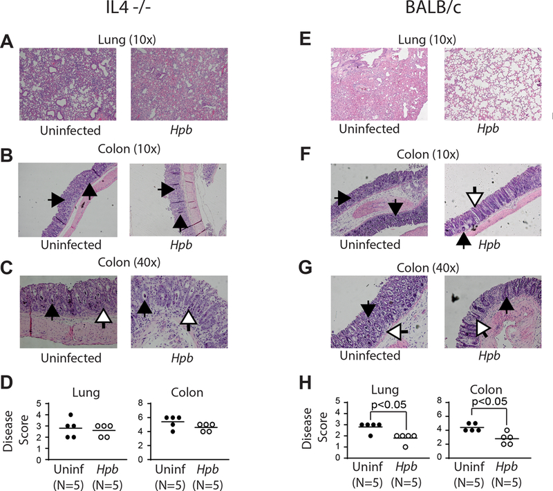
Histopathological analysis of lung (10x magnification) (A, E) and the colon (10x magnification)(B, F), (40x magnification)(C, G) from uninfected and Hpb-infected IL4−/− (A-D) or WT BALB/c (E-H) BMT mice. Organs were harvested 6 days after BMT, tissue preparation and scoring between groups (D, H) was performed as detailed in Methods. Inflammation in the colon was characterized by mononuclear cell infiltrates, apoptotic cells filling crypts (black arrows) and apoptotic bodies (white arrows). Each symbol (dot) is an independent experiment (N) and represents the histopathology score from an individual mouse; bars represent the mean from multiple samples; p values <0.05 between uninfected and Hpb-infected as indicated for each panel; differences between groups determined by unpaired Welch’s t-test.