
Figure 6. MCM2 binds CENP-A and is involved in its maintenance during DNA replication.
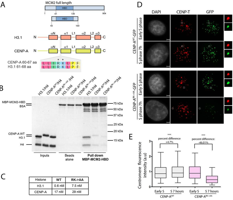
(A) Schematic representation of constructs used in B and C. The CENP-A and H3.1 domain structure is shown. The alignment of an 8 amino acid stretch corresponding to both histones demonstrates the conservation of Arginine 63 and Lysine 64 between the variants. (B) MBP-MCM2-HBD in vitro pull down demonstrating the interaction with indicated histone variants in the wild type and mutant form. (C) Table indicating Kd values measured SPR to assess the strength of interaction between MBP-MCM2-HBD and indicated histone variants in the wild type and mutant form. (D) Representative images of HeLa cells expressing either CENP-AWT-GFP or CENP-ARK->AA-GFP mutant at indicated cell cycle stages. DNA was visualized by DAPI, immunofluorescence for CENP-T is shown in red, expressed CENP-A was detected by GFP signal. Scale bar is 2μm. (E) Quantification of D. The data vas plotted using box-and-whisker plot: 5–95 percentile, n > 5660. The percent change of the levels of centromeric CENP-AWT-GFP and CENP-ARK->AA-GFP forms between experimental time points is indicated, (***) indicates p value <0.0001, statistical significance was calculated using unpaired t-test.