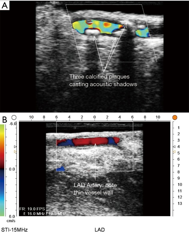
Figure 15.

ECUS of native coronary arteries for determination of best anastomotic site. (A) ECUS of diseased LAD with multiple calcification areas causing acoustic shadows. (B) ECUS of LAD artery in a good location to perform distal anastomosis (ECUS images by Medistim MiraQ system). ECUS, epicardial ultrasound; LAD, left anterior descending artery.
