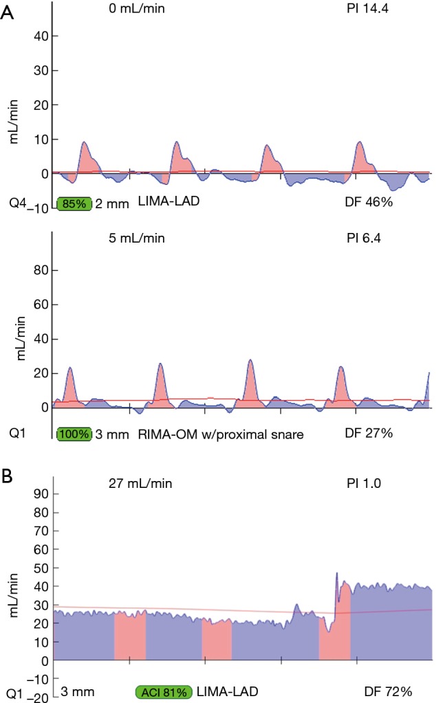
Figure 2.

Transit-time flow measurements of occluded grafts (first two tracings) and the importance of correct probe position on a bypass graft (third tracing). (A) Two examples of ‘true positives’ or occluded grafts with predominant pink spikes of systolic flow and minimal blue waves below the baseline indicating little/no diastolic flow. (B) Example of the mechanism of a ‘false positive’ graft. This reading has caught on screen the changing angle of the probe: on the left side of the image, the probe is not at right angles with the graft and shows lower flow, but when the angle is corrected (right side of image) the flow rises and the PI is good. PI, pulsatility index.
