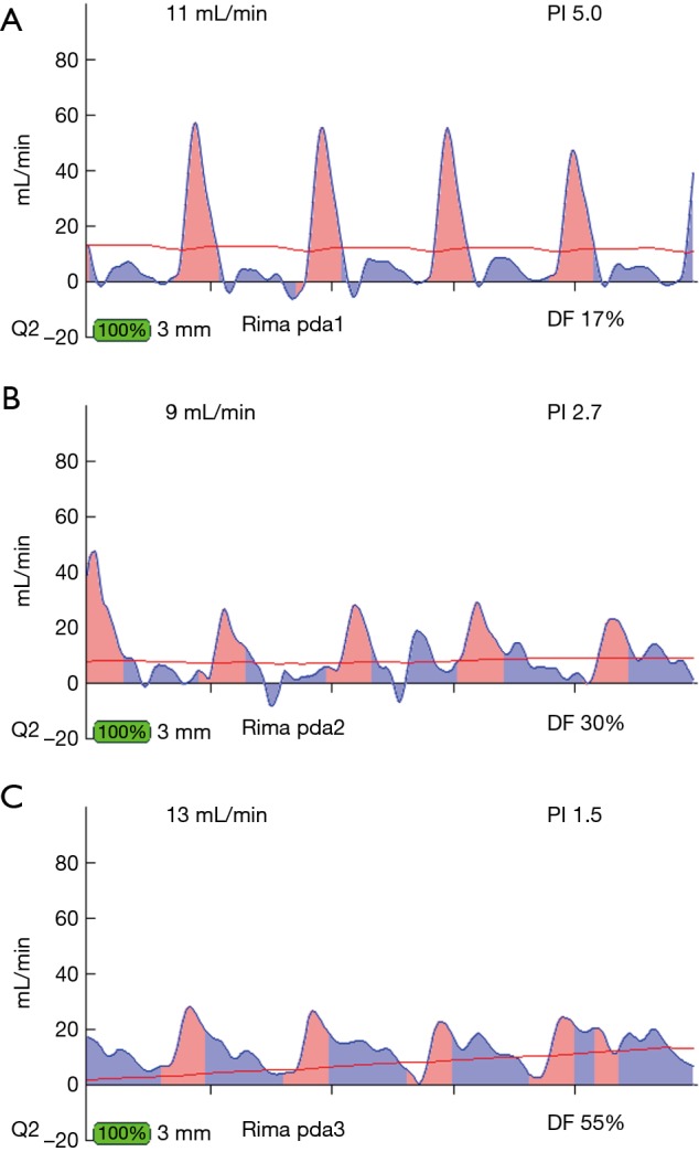
Figure 9.

Different locations of TTFMs on RIMA conduit of RIMA to PDA graft. (A) TTFM of RIMA to PDA close to the proximal end near the subclavian artery. Note the predominant pink spike, as the RIMA in this location is proximate to the systemic circulation. (B) TTFM of the same RIMA to PDA at the midpoint of the conduit. Note that there is more filling in diastole than at the proximal end of the conduit. (C) TTFM of the same RIMA to PDA taken at the distal end of the conduit just ahead of the distal anastomosis. Note that the majority of the filling is in diastole. TTFM, transit-time flow measurement; RIMA, right internal mammary artery; PDA, posterior descending coronary artery.
