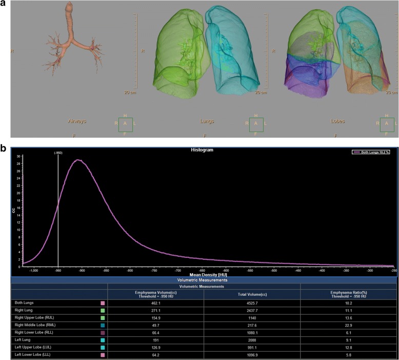
Fig. 1.
Example of the semi-automatic software which was used for emphysema quantification. First, the airways, lungs and lung lobes are segmented (a). Subsequently, a histogram is made which displays the number of voxels with a certain density (b). In this example the percentage of voxels below -950 HU is displayed

