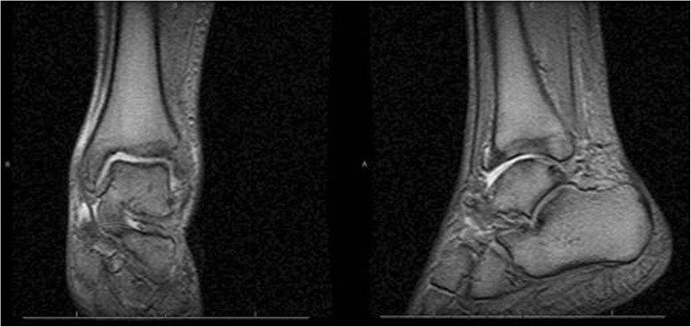
Fig. 1.
Osseous structures: subchondral cysts are present and are described below. Soft tissues: circumferential soft tissue swelling is present, particularly over the malleoli. Articular surfaces: a moderate joint effusion is seen in the tibial talar joint with minimal fluid in the subtalar joint. There is advanced arthrosis of the tibial talar joint with denuding of the articular surface cartilage and subchondral cyst formation in the distal tibia and across the talar dome with subtle mechanical remodeling of the talar dome. Bulky osteophytic ridging is seen anterior distally as well. This bulky osteophytic ridging may be somewhat restrictive in dorsiflexion. There is advanced arthrosis of the tibial talar joint characterized by joint space and bulky anterior osteophytic ridging. Ligaments: thickening of the anterior tibiofibular and anterior talofibular ligaments suggesting residua from prior sprain

