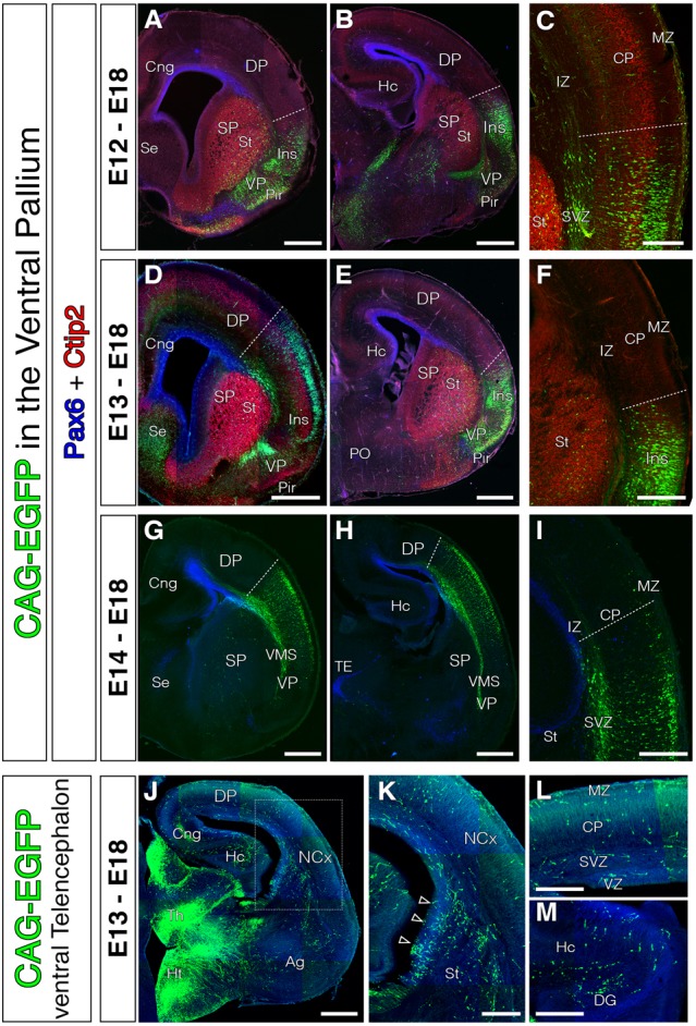
FIGURE 2.

Absence of tangential migration from VP midgestational progenitors to the murine NCx. In all coronal sections, medial is at the left. (A–I) GFP cells derived from the lateral telencephalic wall (comprising SP, VP, LP and in occasions the most lateral DP), electroporated at E12 (A–C, n = 4), E13 (D–F, n = 3), or E14 (G–I, n = 4). We observed no cells that migrated into the DP by the end of neurogenesis (E18); all cells settled within the radial derivatives of the electroporated region (lns, Cl, VP, Pir, VMS, and Ag). Pax6 (blue) and Ctip2 (red) label telencephalic landmarks such as pallial-subpallial boundary, St respectively. (C,F) Dashed lines mark the dorsal limit of the area occupied by the labeled cells along the DP. Very exceptional cells were located slightly beyond this limit, but were not considered tangential cells given their proximity to the electroporation area and their radial morphology – lns boundary. (J–L) Electroporations at E13 on other telencephalic regions (including the ventral SP) labeled large populations of tangential migratory interneurons. (K) High power view of the region indicated with rectangle in J. (L–M) High power views from the dorsal NCx and hippocampus (Hc) showing the horizontally oriented tangential interneurons. DAPI counterstain in blue in (J–M). Scale bars represent 500 μm and 250 μm in (A–M).
