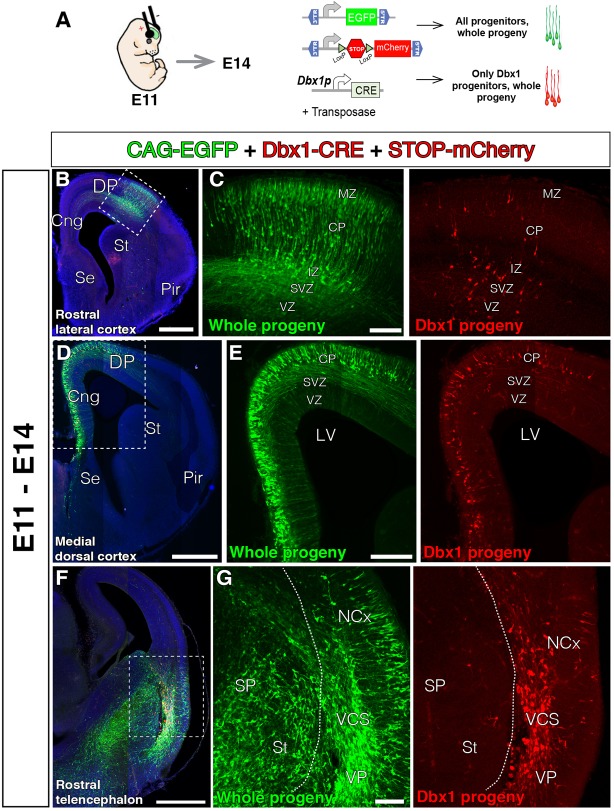
FIGURE 3.
The lineage of early local DP Dbx1-expressing progenitors. In all coronal sections, medial at the left. DAPI counterstain in blue. (A) Electroporation paradigm at E11 (n = 4). The DNA cocktail simultaneously labels the lineage of any progenitors (green) and of Dbx1-expressing progenitors (red). The location of the labeled cells is analyzed and documented 3 days later. (B–E) Dbx1-expressing progenitors generate neurons that migrate radially to the cortical plate (CP), both at the DP-derived NCx (B, power view in C) and the MP-derived cingulate cortex (D, power view in E). Dbx1-derived neurons represented a small fraction of the total electroporated cells. (F,G) Power views from the lateral telencephalon. Some dorsal pallial cells are derived from Dbx1-expressing progenitors (red). At the subpallium (SP), many cells were electroporated (EGFP, green in F,G), but no cells expressed the red protein, suggesting Dbx1 enhancer is selectively active in scattered pallial progenitors. Dashed line marks the pallial-subpallial boundary. Scale bars represent 500 μm; 200 μm; and 100 μm in (B–G).

