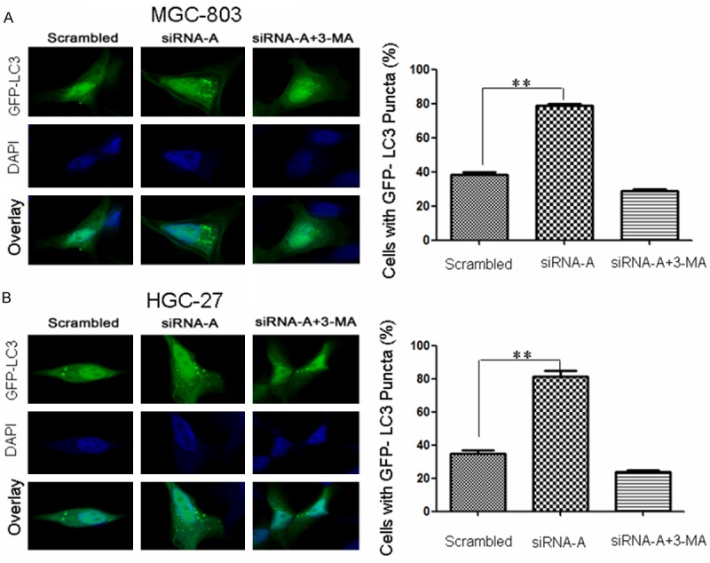
Figure 1.

URI siRNA-A transfection enhanced GFP-LC3 puncta aggregation in gastric cancer cells. Cells were transfected with a plasmid encoding EGFP-LC3. Autophagosomes were visualized by the presence of GFP-LC3 puncta (green). DAPI staining (blue) was used to detect nuclei. GFP-LC3 puncta were observed by confocal microscopic analysis of MGC-803 (A) and HGC-27 (B) URI siRNA-A-transfected cells with or without 3-MA treatment. The percentage of cells positive for GFP-LC3 puncta (containing five or more GFP-LC3 punctate dots per cell) is shown (right panel). Data are presented as the mean ± SD of three independent experiments (100 cells were counted for each experiment). **P < 0.01.
