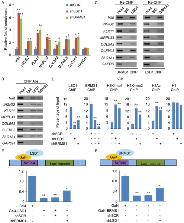
Figure 4.
BRMS1 and LSD1 coregulate the expression of VIM. A. qChIP analysis of the indicated genes in MCF-7 cells. Results are represented as fold change over the GAPDH control. Error bars represent mean ± SD for three independent experiments. *P < 0.05 and **P < 0.01 (two-tailed t-test). B. ChIP analysis of the indicated genes in MCF-7 cells by conventional DNA electrophoresis. IgG served as a negative control. C. Re-ChIP analysis of the seven selected co-targets in MCF-7 cells with antibodies against BRMS1 and LSD1 using conventional DNA electrophoresis. IgG served as a negative control. D. qChIP analysis of the recruitment of the indicated proteins on the VIM promoter in MCF-7 cells after infection with control lentivirus-mediated shRNA, or shRNAs-targeting BRMS1 or LSD1. Purified rabbit IgG was used as a negative control. Error bars represent mean ± SD of three independent experiments. *P < 0.05 and **P < 0.01 (two-tailed t-test). E and F. The control vector (containing Gal4-DBD only), Gal-4-LSD1, or Gal4-BRMS1 constructs was prepared and transfected alone or with the indicated specific lentivirus-mediated shRNAs into MCF-7 cells stably expressing Gal4-UAS reporter (MCF-7-Gal4-Luc cells). Gal4 luciferase reporter activity was measured. *P < 0.05 and **P < 0.01 (two-tailed t-test).

