Figure 3.
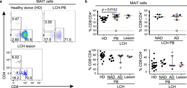
MAIT cell subsets in LCH. (a) Gating strategy for identifying CD8 and CD4 cells in MAIT cells from the peripheral blood from healthy donors (top left) and LCH patients (PB) (top right), and in lesional MAIT cells from patients with LCH (bottom left). (b) Proportions of CD4+, CD8+, and CD8−CD4− cells in MAIT cells from the peripheral blood from healthy donors (HD), patients with non-active LCH (PB-NAD) and active LCH (PB-AD) and in lesional MAIT cells from patients with active LCH. Kruskal-Wallis tests with Dunn’s multiple comparisons were conducted for (b) except where only two groups were compared (Mann Whitney test), error bars indicate median + interquartile range, 10−3 on logarithmic scale indicates ‘undetectable’.

