Figure 1.

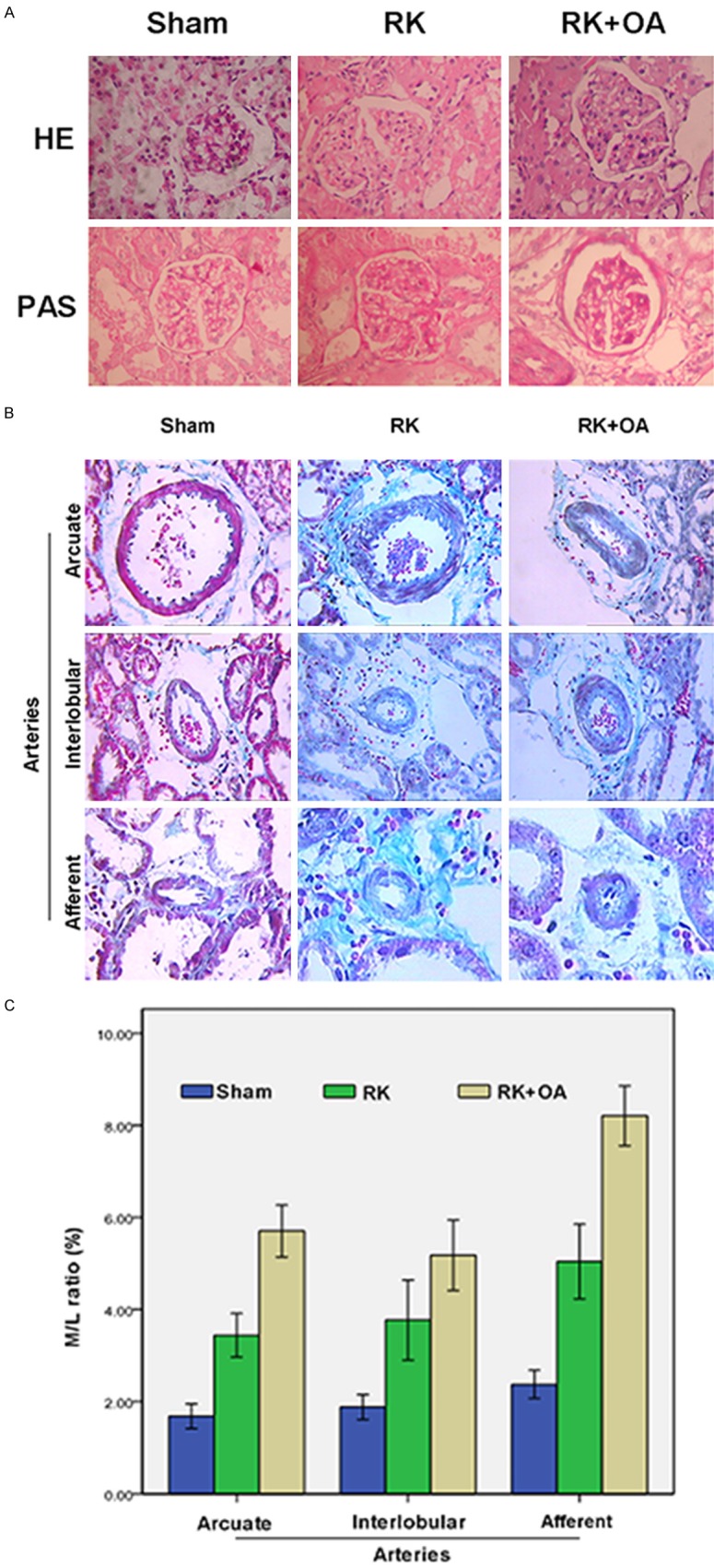
The morphology of glomeruli and preglomerular arteries. A: Histologic examinations were performed after H&E staining (upper panel) and PAS staining (lower panel) to observe the morphology of glomeruli in the Sham, RK, and RK + OA groups. OA aggravated renal damage in RK rats. B: Representative images of arcuate arteries (×200), interlobular arteries (×200), and afferent arteries (×400). C: The wall thickness of preglomerular arteries was evaluated by calculating the M/L ratio. OA promoted periarterial adventitial fibrosis and thickening of the preglomerular artery in RK rats
