
Figure 2.
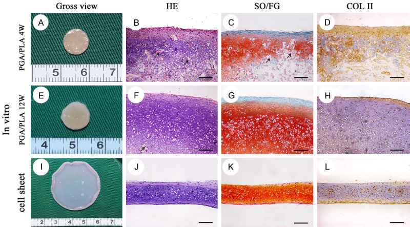
Gross view and histological staining of the in-vitro pre-cultivated cartilage cell sheet and chondrocytes-PGA/PLA composition. Gross view of chondrocytes-PGA/PLA composition in-vitro pre-cultivation for 4 w (A), 12 w (E) and cartilage cell sheet chondrogenic cultured for 3 w (I). The in-vitro pre-cultivated construct showed great many alive chondrocytes with typical lacunae (B, F, G; HE), strong positive staining of safranin O/fast green (C, G, K; SO/FG) and type II collagen (D, H, L; COL II). A number of PGA/PLA fiber still could be detected (black arrow) in histological staining section (B, C, F). Scaffold-free cartilage cell sheet was membrane without any foreign material residual in histological staining section (I-L). scale bar = 100 um.