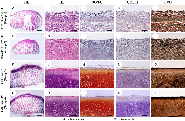
Figure 7.
Histological and immunohistochemical staining of the in-vivo regenerated cartilage-phase construct in different groups. In group 1 (A-E), confused loose connective tissue was detected. In group 2 (F-J), the regenerated construct displayed disorderly dense connective tissue with uncertain structure. Group 3 and group 4 showed alive chondrocytes, typical lacunae (K, L, P, Q; HE) with strong positive staining of safranin O/fast green (M, R; SO/FG), type II collagen (N, S; COL II) and elastic fibre (O, T; EVG). Scale bar = 1000 um (A, F, K, P), scale bar = 100 um (B-E, G-J, L-O, Q-T).

