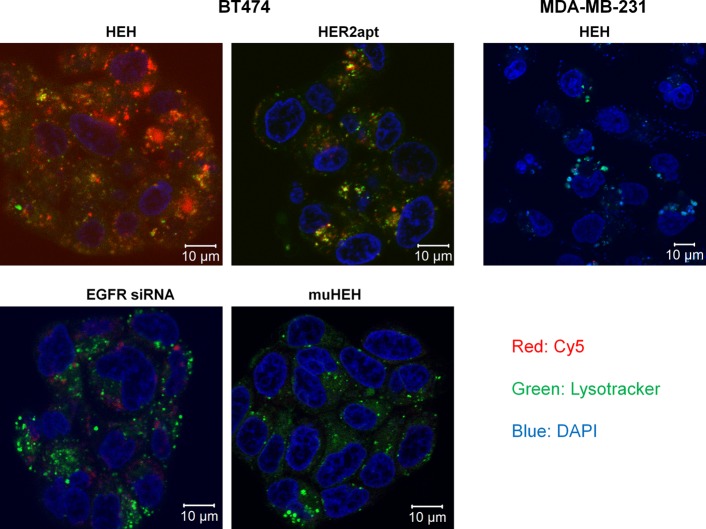
Figure 3.
Detection of HEH internalization by Z-stack confocal microscopy. Cy5-labeled HEH, muHEH, HER2 aptamer, or EGFR siRNA was added into BT474 cells for 12 h at 37 °C. Lysotracker Green and DAPI were added into cells at the same time as the chimeras. LysoTracker Green was used to show lysosomes and endosomes. DAPI (blue) was used to display nucleus. Confocal laser scanning microscopy with z-stack was performed to show cell binding and internalization.

