Fig. 4.
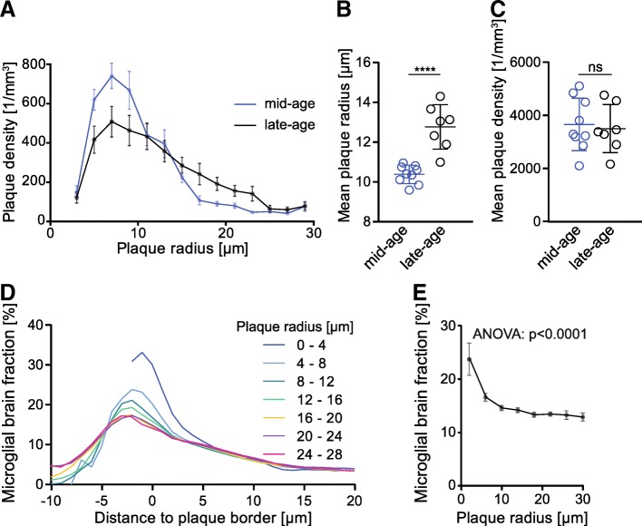
Molecular elucidation of in vivo μPET findings by terminal immunohistochemistry. a Frequency distribution of plaque radii in mid-aged (11.4 to 12.7 months) and late-aged (13.6 to 15.3 months) APP-SL70 mice. The mean plaque radius (b) is significantly higher in the late-aged cohort when compared to mid-aged APP-SL70 mice (p < 0.0001, two-tailed Student’s t test), whereas the plaque density (c) did not indicate changes during aging > 12 months in APP-SL70 mice (p = 0.746, two-tailed Student's t test). d Correlation of microglial brain fraction with distance to plaque border and plaque size. Each profile represents the change of microglial brain fraction with distance to the border of plaques with defined radius. e Microglial brain fraction in the vicinity to the plaque border (radius 1 μm) decreased significantly with increasing plaque radius (one-way ANOVA, F(5,16) = 11.87, p < 0.0001). Data presented as mean ± SEM; n = 7–9 mice

