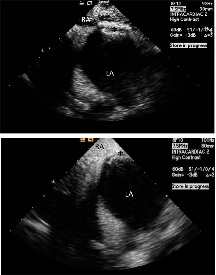
Figure 11.

Intracardiac echocardiogram (ICE) image of a released Amplatzer PFO Occluder (top) (*) and Gore Cardioform Septal Occluder (bottom) (*). Agitated saline is seen in the right atrium (RA), but not in the left atrium (LA), confirming the absence of right to left shunt after device deployment.
