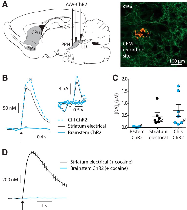
Figure 2.

Light activation of striatal cholinergic brainstem afferents with brief stimuli does not reveal dopamine release. A, Left, cartoon indicating PPN/LDT injections sites, and right, recording site labeled with red FluoSpheres in an area of cholinergic brainstem innervation indicated by ChR2-eYFP fluorescence. B, Example profiles of [DA]o (μm) versus time evoked by either light activation of ChIs (ChI ChR2, blue dashed, 1p), electrical stimulation of striatum (black solid line, 1p), or light activation of brainstem cholinergic afferents (brainstem ChR2, blue solid line, 10p 10 Hz). Inset, corresponding voltammograms for DA following each activation type for site indicated in A, at time point indicated on profiles by gray circles. C, Peak evoked [DA]o (μm) for each recording site and stimulation methods, with mean ± SEM indicated. Arrows indicate data points and sites shown in B. D, Mean profiles of current detected at DA oxidation potential (± SEM) versus time evoked by striatal electrical stimulation (1p; black) or light stimulation of ChR2-expressing cholinergic brainstem afferents (10p 10 Hz; blue) in the presence of cocaine (5 μm; n = 10 observations from 3 sites).
