Figure 3.
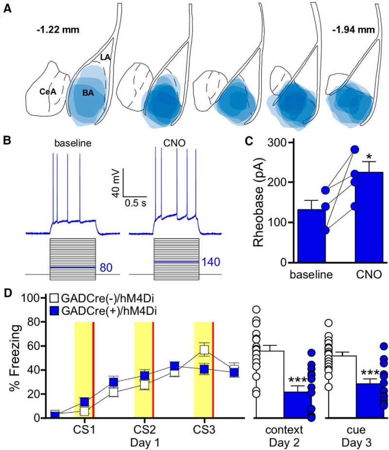
Chemogenetic inhibition of BA GABA neurons impairs auditory fear conditioning. A, Schematic summarizing the distribution of hM4Di-mCherry fluorescence in the BA (coronal view, spanning –1.22mm to –1.94mm posterior from bregma) of GADCre(+)/hM4Di mice evaluated in D. B, CNO-induced inhibition of an hM4Di-expressing BA GABA neuron. Rheobase was measured before (baseline) and after application of CNO (10 μm). The current-step protocol is depicted below the traces. The first step to elicit spiking is highlighted in blue; the trace shown is the response to the denoted current step. C, Rheobase summary for hM4Di-expressing BA GABA neurons, before and after CNO application (t(4)=3.2, *p = 0.033). Each experiment is shown as connected circles (n = 5). D, Impact of BA GABA neuron inhibition on fear learning. GADCre(+)/hM4Di (blue) and GADCre(−)/hM4Di (white) mice were trained with a 3 CS/3 US paradigm. CNO (2 mg/kg, i.p.) was given 30 min before training. Freezing during training is shown on the left. The yellow bars denote CS presentations and the red bars indicate US presentations. There was no main effect of genotype (F(1,174)=0.1, p = 0.71). The plots on the right show freezing during context (t(29)=4.9, ***p < 0.001) and cue (t(29)=4.5,***p < 0.001) tests. Error bars represent the mean ± SEM, with dots next to the bars denoting individual data points (n = 6/8–9/8 males/females group).

