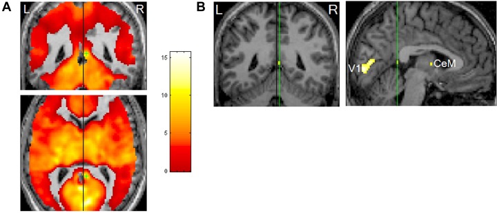
FIGURE 4.
Rapid eye movements (REM)-locked activation in retrosplenial cortex (RSC). (A) Upper panel shows coronal view, lower axial view. Black lines indicate the mid-sagittal plane. Green crosshair is at the right RSC peak activity point (Talairach coordinates 4, –46, 12, t = 10.5). Threshold has been reduced to uncorrected P < 0.5 (t = 0) to illustrate the failure to detect significant responses in the RSC on the left hemisphere, which contrasts with the adjacent robust activation in the right hemisphere. (B) Left panel shows a coronal view and right sagittal view. The black line on the coronal view indicates the mid-sagittal plane. Green lines pass the right RSC peak activity point and show location of the other views. Thresholded higher at corrected P < 0.00005 (t = 10.1). V1, primary visual cortex; CeM, central medial thalamic nucleus (a part of intralaminar/midline group). Adapted from Hong et al. (2009) with the permission from Hong.

